Plenary Speakers
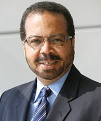
Dr. Roderic Pettigrew
NIH/NIBIB, USA
Title: Advancing Precision Medicine through Biomedical Imaging
Tuesday April 29, 2014,13:40-14:30
Roderic I. Pettigrew, MD, PhD, is the first Director of the National Institute of Biomedical Imaging and Bioengineering at the NIH. Prior to his appointment at the NIH, he was Professor of Radiology, Medicine (Cardiology) at Emory University and Bioengineering at the Georgia Institute of Technology and Director of the Emory Center for MR Research, Emory University School of Medicine, Atlanta, Georgia.
Dr. Pettigrew is known for his pioneering work at Emory University involving four-dimensional imaging of the cardiovascular system using magnetic resonance (MRI). Dr. Pettigrew graduated cum laude from Morehouse College with a B.S. in Physics, where he was a Merrill Scholar. He earned an M.S. in Nuclear Science and Engineering from Rennselear Polytechnic Institute; and a PhD in Applied Radiation Physics from the Massachusetts Institute of Technology, where he was a Whitaker Harvard-MIT Health Sciences Scholar. Subsequently, he received an MD from the University of Miami School of Medicine in an accelerated two-year program, did an internship and residency in internal medicine at Emory University and completed a residency in nuclear medicine at the University of California, San Diego. Dr. Pettigrew then spent a year as a clinical research scientist with Picker International, the first manufacturer of MRI equipment, where he helped develop their first cardiac imaging technology. In 1985, he joined Emory as a Robert Wood Johnson Foundation Fellow with an interest in non-invasive cardiac imaging. His current research focuses on integrated imaging and predictive biomechanical modeling of coronary atherosclerotic disease.
Dr. Pettigrew’s awards include membership in Phi Beta Kappa, the Bennie Award (Benjamin E. Mays) for Achievement, and being named the Most Distinguished Alumnus of the University of Miami (1990). He was the Radiological Society of North America’s 75th Diamond Jubilee Eugene P. Pendergrass New Horizons Lecturer. He is also the recipient of the Herbert Nickens Award of the ABC, the Pritzker Distinguished Achievement Award of the Biomedical Engineering Society, and the Distinguished Service Award of the National Medical Association. He has been elected to membership in two components of the US National Academies: the Institute of Medicine, and the National Academy of Engineering.
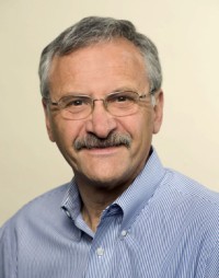
Dr. Norbert J. Pelc
Stanford University, USA
Title: Promising Directions for CT Technology Development
Wednesday, 30 April, 2014, 10:00-10:50am
Norbert J. Pelc received his bachelor’s degree in 1974, and masters and doctoral degrees in Radiological Physics at Harvard in 1976 and 1979. Dr. Pelc worked in industry from 1978 until 1990 where he was instrumental in the development of CT, MRI, and digital radiography. He joined Stanford in 1990. He is Professor of Bioengineering and Radiology, and Electrical Engineering (by courtesy), and Chair of Bioengineering.
Dr. Pelc’s research interests are in diagnostic imaging, especially MRI and CT. His current research focuses on advanced CT system design and reconstruction methods, and in the development of new applications. He has authored 185 papers, over 300 presentations, and 88 US patents. He is a Fellow of the AAPM, of the International Society of Magnetic Resonance in Medicine (ISMRM), the American Institute of Medical and Biological Engineering (AIMBE), and the Council on Cardiovascular Radiology of the American Heart Association. He was awarded the Edith H. Quimby Award by the AAPM and the Outstanding Researcher Award by the RSNA. He was a member of the first Advisory Council of the National Institute of Biomedical Imaging and Bioengineering (NIBIB) and is currently a member of the Council of Councils of the NIH. In 2012 was elected to the National Academy of Engineering in recognition of his contributions to the development of CT and MRI.
Abstract: Computed Tomography (CT) has made enormous technical advances since its introduction into clinical use. The engineering improvements have in turn led to important clinical applications and large impact in patient care. This paper reviews the technology development trends in Computed Tomography since its introduction and uses these trends to help illuminate likely future progress. Advances have been motivated by the needs of new clinical applications, and recently also by the desire to reduce radiation dose to the patient. Scanning speed, measured in image data points or raw data points acquired per second, has grown roughly exponentially since the 1970’s. The trend, even in the component technologies that drive it, show no little of slowing. Spatial resolution showed strong improvement in early years but has plateaued due to the difficulty of improving resolution with current technology. Direct conversion photon counting detectors could provide a significant improvement in resolution, and also in other ways by eliminating the degradation from electronic noise, by increasing the geometric efficiency, and by providing spectral information. However, increasing the count rate capability and achieving good energy response are a significant challenge. Advanced reconstruction methods have made strong impact in recent years, and this is expected to continue. Concerns about radiation dose have driven technology in recent years and this will continue. More precise control of the x-ray illumination field can reduce the radiation dose needed to obtain the needed image quality and also facilitate introduction of photon counting detectors. Overall, the prediction is that significant further improvements in speed, spatial resolution and dose efficiency can be expected in the next decade.
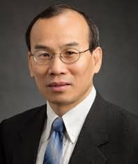
Dr. Zhi-Pei Liang
University of Illinois at Urbana-Champaign, USA
Title: MR Metabolic Imaging: A Path to High Resolution through Subspaces
Thursday, 1 May, 2014, 10:05am-10:55am
Zhi-Pei Liang received his Ph.D. degree in Biomedical Engineering from Case Western Reserve University in 1989. He subsequently joined the University of Illinois at Urbana-Champaign. He is currently Franklin W. Woeltge Professor of Electrical and Computer Engineering and Co-chair of the Integrative Imaging Theme of the Beckman Institute for Advanced Science and Technology. Dr. Liang’s research covers image formation theory, algorithms, and biomedical applications. For his research contributions, he has received a number of prestigious awards, including the Sylvia Sorkin Greenfield Award (Medical Physics, 1990), an NSF CAREER Award (1995), and the Otto Schmitt Award from the International Federation for Medical and Biological Engineering (2012). Dr. Liang is a Fellow of AIMBE (2005), IEEE (2006), and ISMRM (2010), and was elected to the International Academy of Medical and Biological Engineering in 2012. He served as President of IEEE Engineering in Medicine and Biology Society from 2011-2012.
Abstract: MR spectroscopic imaging (MRSI or spatially-resolved MR spectroscopy) has been recognized as a powerful tool for noninvasive metabolic studies of biological systems but clinical and research applications of this technology have been developing very slowly. Conventional MRSI methods represent the desired spatiospectral function as a vector in a very high-dimensional space. As a result, the number of spatiospectral encodings required for decoding (or image reconstruction) can be huge, resulting in long data acquisition time, or poor spatial resolution, or low signal-to-noise ratio. It can be justified that the spatiospectral functions of real biological systems reside in a very low-dimensional subspace. This property can be effectively utilized to accelerate MRSI experiments and achieve high resolution. This talk will discuss our recent advances in this exciting area of research.
Dr. Brian W. Pogue
Dartmouth College, USA
Title: Optical Molecular Imaging in Humans
Friday, 2 May, 2014, 10:10am-11:05am
Brian W. Pogue, Ph.D. is Professor of Engineering, Physics & Astronomy, at Dartmouth College, and Adjunct Professor of Surgery at the Geisel School of Medicine. He has a doctoral degree in Medical Physics from McMaster University in Canada, and maintains a visiting research position at the Massachusetts General Hospital. His research is in optical imaging systems, with a focus on molecular and structural imaging of cancer for surgical guidance and imaging radiation therapy. He has published 240 peer-reviewed papers. His research is funded by several grants from the National Cancer Institute USA and he is a fellow of the Optical Society of America. He currently serves as chair of the Biomedical Imaging Technology A (BMIT-A) Study Section, and is an editorial board member for the Journal of Biomedical Optics, Medical Physics and Physics in Medicine and Biology.
Abstract: Optical signals provide the ability to quantify molecular or transport features of tissue, with sensitivity down to nanoMolar concentrations in vivo. This extreme molecular sensitivity is only avauilable through nuclear medicine or optical tracers, and while nuclear medicine tracers provide optimal means for diagnostic imaging, optical tracers are optimal for surgical or interventional guidance. Many optical tracers for cancer have been studied in pre-clinical animal models showing the ability to localize due to compartmentalization, retention, binding or internalization. These can be used for diagnostic imaging or interventional guidance, with the latter application being perhaps more financially viable in the near term. Clinical trials using non-specific fluorescent molecules will be reviewed, including indocyanine green, fluorescein, methylene blue, and protoporphyrin IX, and some ongoing and future plans for molecular-targeted and activable tracers outlined. The imaging of binding in and molecular expression in vivo can be achieved and the pathways for seeing this enter the healthcare world will be discussed in both diagnostic imaging to guide drug development as well as interventional imaging for procedure guidance.

