Special Session
Title 1: Advances in Computer-Aided Histopathology
Session organizers: Carolina Wählby, Ingrid Carlbom, Alexandra Pacureanu
Tuesday, 29 April – 16:20-18:00 – Room R&F 3
Session Description:
Detection, diagnosis, and severity-grading of cancer are traditionally based on the visual examination under a microscope of histopathological tissue samples. The current transition from visual examination of glass slides under microscopes to whole slide scanners and computer-aided image analysis, i.e., digital pathology, holds the promise of more objective cancer grading that will lead to better prognostication while at the same time reducing the pathologist’s workload. In this session we will review the historical development of digital histopathology, present novel methods that will lead to automatic and objective cancer grading in clinical practice, and discuss how recent advances in spatial mapping of biomedical markers may improve prognostication in the future.
Biography of the organizers:
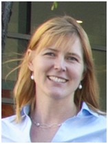 Carolina Wählby is Principal Investigator at the Imaging Platform at the Broad Institute of Harvard and MIT, Cambridge, MA, and Associate Professor at the Div. of Visual Information and Interaction, Dept. of Information Technology, Uppsala University, Sweden. Her research group is partly funded by SciLifeLab Sweden, and includes algorithm development for robust detection and feature extraction from primarily microscopy images with applications in the biomedical field, ranging from image based RNA sequencing to high-throughput screening assays using primary cell cultures from human tumors and model organisms such as C. elegans. Dr. Wählby earned her PhD in Digital Image Analysis at the Centre for Image Analysis at Uppsala University in 2003, and carried out post-doctoral research at the Dept. of Genetics and Pathology at the same university. She established her own research group at the Centre for Image Analysis at Uppsala University in 2007, and was recruited as Principal Investigator at the Imaging Platform at the Broad Institute of Harvard and MIT in 2009. She was selected as ‘strategic recruitment’ for SciLifeLab Sweden in 2011 and has received recognition and research funding from the National Institutes of Health (NIH R01), the Swedish Research Council (Young investigator grant) and SciLifeLab Sweden.
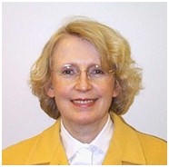 Ingrid Carlbom is Professor at the Division of Visual Information and Interaction, Department of Information Technology, Uppsala University, Sweden, where her research includes automatic malignancy grading of prostate cancer, and haptic interaction with applications to cranio-maxillofacial surgery planning. Previously she was Director of Visual Communications Research at Bell Laboratories, Lucent Technologies, Murray Hill, NJ; Manager of Visualization Research at Digital Equipment Corporation’s Cambridge Research Lab in Massachusetts; and Program Leader for the Graphical Modeling Program at Schlumberger-Doll Research, Ridgefield, CT.
Dr. Carlbom received a Ph.D. in computer science from Brown University. She has been active in SIGRAPH as a director and as the Chair of the SIGGRAPH Advisory Board. She has served on the editorial boards of IEEE Computer Graphics and Applications, IEEE Transactions on Visualization and Computer Graphics, and Computers & Graphics, and has served as Co-Editor-in-Chief of Graphical Models. She was a member of the Advisory Board for the NSF/ARPA Science and Technology Center for Computer Graphics and Scientific Visualization. From 2004 through 2010 she has chaired the board of the Graduate School in Mathematics and Computing at Uppsala University. She received a Doctor of Philosophy Honoris Causa from Uppsala University, and a Distinguished Graduate School Alumna Award from Brown University.
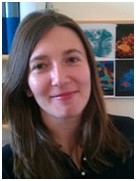 Alexandra Pacureanu earned her PhD in Electrical Engineering from INSA de Lyon, Université de Lyon, France, in 2012. Her thesis focused on nanoscale X-ray tomographic imaging with a synchrotron radiation source and image analysis applied in biomedicine, for which she was awarded the 2012 IEEE EMBS French section, SFGBM and SticSanté Innovation price. She is now a postdoctoral research fellow in the Quantitative Microscopy group at the Centre for Image Analysis and Science for Life Laboratory, Uppsala University, Sweden.
Session organizers: Françoise Peyrin, Atsushi Momose, Max Langer
Wednesday, 30 April -11:00-12:40 – Room R&F3
Session Description:
X-ray phase imaging associated to different experimental systems is receiving an increasing attention for applications going from microscopy to clinical imaging. Compared to X-ray absorption imaging this technique offers potentially a three-orders of magnitude increase in sensitivity and has a strong interest for resolving small density variations or imaging soft tissue. This technique is generally associated to a so-called phase retrieval problem. The aim of this session, is to provide an overview of phase imaging techniques and of the associated inverse problem that are of interest for ISBI attendants.
Biography of the organizers:
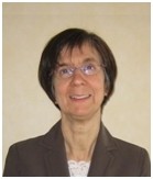 Françoise Peyrin is Director of Research at INSERM (National Institute for Health and Medical Research) in Lyon, France. She is leading a group on « Tomographic imaging and Radiotherapy » in the CREATIS Laboratory (INSERM U1044, CNRS 5220, INSA-Lyon, Université Lyon I, University of Lyon) specialized in medical imaging and gathering about 180 staff. She received her PhD in Computer Sciences and her “Docteur ès Sciences” degree from INSA-Lyon and Université Lyon I, respectively in 1982, and 1990. She has been Assistant Professor at INSA-Lyon from 1981 to 1987, and since then a Researcher at INSERM. Since 1995, she has been a scientific collaborator at the ESRF (European Synchrotron Radiation Facility), Grenoble, France. Her research interest is in 3D biomedical imaging particularly in X-ray tomography, tomographic image reconstruction, image analysis and wavelet based methods. She is particularly developing new image analysis methods for the characterization of bone tissue at the micro and nano scales. She is the author of 150 peer-reviewed papers, 18 books chapters and more than 300 conference papers (h-index:31). Since 2007, she has been in the committee of the French GDR Stic Sante that she is currently leading. Since 2003, she has been serving as a member of the IEEE Bio Imaging and Signal Processing Technical committee. Since 2012, she is leading the LabEx (Laboratory of Excellence) PRIMES (Physique Radiobiologie Imagerie Médicale et Simulation) at Université de Lyon.
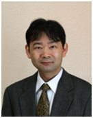 Atsushi Momose is a professor at IMRAM (Institute of Multidisciplinary Research for Advanced Materials), Tohoku University, Japan. He received his PhD degree of Engineering from The University of Tokyo in 1996. After finishing the master course of the Department of Applied Physics, Graduate School of Engineering, The University of Tokyo in 1987, he joined Advanced Research Laboratory, Hitachi, Ltd.. He has been studying methodology with synchrotron radiation mainly at Japanese synchrotron radiation facilities, the Photon Factory, KEK and SPring-8. From 1997 to 1998, he stayed at the ESRF (European Synchrotron Radiation Facility), Grenoble, France as a visitor scientist. He is known especially by his studies on X-ray phase imaging performed since 1990s. In 1999, he moved to The University of Tokyo, as an associate professor, and opened up a new frontier of X-ray phase imaging; that is X-ray grating interferometry, enabling practical use of X-ray phase imaging outside of synchrotron facilities.
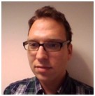 Max Langer is a CNRS researcher at the University of Lyon, France, in the CREATIS laboratory and a visiting scientist at the European Synchrotron Radiation Facility, Grenoble, France. His main research interests concerns inverse problems in X-ray phase contrast imaging, tomographic reconstruction and high resolution X-ray imaging of bone ultrastructure. He received his PhD degree in electrical engineering in 2008 from the INSA-Lyon for his work on phase retrieval in the Fresnel region for hard X-ray tomography, and his MSc degree in biomedical engineering in 2004 from Linköping University, Sweden, for his work on the design of fast multidimensional filters using genetic algorithms. He has authored more than 80 peer reviewed articles He received a French scientific excellence bonus for the period 2014-2017.
Title 3: Computational Methods for Optical Imaging and Tomography
Session organizers: Yujie Lu, Gultekin Gulsen
Wednesday, 30 April -14:00-15:40 – Room R&F3
Session Description:
Optical imaging has been playing an important role in preclinical research and is being translated to the clinical for disease diagnosis and therapy. With the development of new molecular optical imaging techniques, a series of challenging problems needs to be solved to improve and optimize the imaging performance, including the modeling of interaction between the optical photons and different tissue types; tomographic reconstruction and optimization algorithms; and signal processing as well as image pre- and post-processing techniques. The proposed special session will focus on optical imaging and tomography field and report the latest progress in modeling, image reconstruction and processing of all optical imaging modalities.
Biography of the organizers:
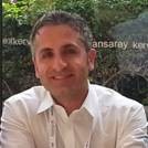 Gultekin Gulsen is an Associate Professor of Radiology and Biomedical Engineering at University of California, Irvine (UCI), with a primary appointment at Center for Functional Onco-Imaging. Dr. Gulsen received his Ph.D. degree in Applied Physics from Bosphorus University in Istanbul, Turkey. For more than a decade, his team has been spending effort to achieve molecular imaging with high resolution and quantitative accuracy by mainly integrating laser-based optical molecular imaging with anatomic imaging modalities such as MRI and X-ray CT. For example, his team has developed a gantry-based combined X-ray CT and Fluorescence Tomography system for animal imaging. This system is unique in that both X-ray CT and optical imaging systems are on the same gantry rotating around the animal and capable of revealing cross-sectional background absorption map and anatomic images to obtain quantitatively correct fluorophore concentration maps. Another hybrid system that is being developed in his lab is a combined MRI-Diffuse Optical Tomography scanner for small animal imaging in absorbance & fluorescence modes. Recently, Dr. Gulsen’s team started building combined MRI and nuclear imaging systems for breast cancer such as MRI-SPECT & MRI-PET. His research is funded by awards from United States National Institutes of Health, Department of Defense Breast Cancer Program, California Breast Cancer Research Program, and Susan G. Komen foundation. He is a member of Institute of Electrical and Electronics Engineers (IEEE), Biomedical Engineering Society (BMES), Optical Society of America (OSA), and SPIE. He serves in the Review Editorial Board of Frontiers in Biomedical Physics.
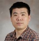 Yujie Lu, an assistant professor in Center for Molecular Imaging (CMI), Institute of Molecular Medicine, University of Texas Health Science Center at Houston. He received his Ph.D. degree from Institute of Automation, Chinese Academy of Science in 2007. He completed his two-year postdoctoral training in Crump Institute for Molecular Imaging, UCLA, where he was devoted to developing a novel multimodal Optical-PET (OPET) imaging system. Since 2009, he is Tomography Core Leader in CMI to build a trimodal (Optical/PET/CT) tomography imaging system in collaboration with Siemens and to seek relevant biological application and clinical translation. His research interests include theoretical and numerical methods for inverse problem in biomedical imaging modality especially optical tomography, small animal multimodal imaging system, and preclinical and clinical translational applications. He has authored/co-authored about 50 peer-reviewed journal and conference papers. Two papers were selected as “Featured Article” by Physics in Medicine and Biology and one paper was selected as “Highlights of 2010” by the same journal. He serves as an Editorial Board Member for the Scientific World Journal and a Guest Editor for Computational and Mathematical Methods in Medicine.
Title 4: Laser Speckle Imaging and Biomedical Applications
Session organizers: Shanbao Tong, Bernard Choi
Wednesday, 30 April – 16:10-17:50 – Room R&F3
Session Description:
Laser Speckle Imaging is a 2D, noninvasive blood flow imaging technique, and can provide both the structural information and functional responses in neurovascular systems. LSI now has been used in a wide range of biomedical engineering pre-clinical research and biomedical applications, such as stroke, angiogenesis, retinal system and etc. In this special session, we are going to invite 5-6 experts who have been actively working in this area, either in the techniques of the LSI or exploring the potential biomedical applications.
Biography of the organizers:
Bernard Choi is an Associate Professor of Biomedical Engineering and Surgery at University of California, Irvine (UCI), with a primary appointment at Beckman Laser Institute and an affiliated appointment in the Edwards Lifesciences Center for Advanced Cardiovascular Technology. Dr. Choi received a B.S. from Northwestern University and M.S.E. and Ph.D. degrees with The University of Texas at Austin, all in Biomedical Engineering. His current research is in the field of vascular biophotonics, with current emphases in optical hemodynamic monitoring, microvascular dynamics of tissue in normal and proliferative states, and optical clearing. Dr. Choi is a co-author on 88 peer-reviewed journal publications. His research is funded by awards from Air Force Office of Scientific Research and the United States National Institutes of Health. He has received several UCI teaching awards, including Excellence in Undergraduate Education (2013), Biomedical Engineering Faculty of the Year (2009, 2010, 2013), and the Chancellor’s Award for Excellence in Fostering Undergraduate Research (2007). He is a member of American Society for Engineering Education (ASEE), American Society for Laser Medicine and Surgery (ASLMS), Biomedical Engineering Society (BMES), the Microcirculatory Society, Optical Society of America (OSA), and SPIE.
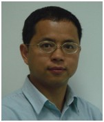 Shanbao Tong received the B.S. degree in radio technology from Xi’an Jiao Tong University, Xi’an, China, in 1995, the M.S. degree in turbine machine engineering, and the Ph.D. degree in biomedical engineering from Shanghai Jiao Tong University, Shanghai, China, in 1998 and 2002, respectively. From 2000 to 2001, he was a Research Trainee in the Biomedical Instrumentation Laboratory, Biomedical Engineering Department, Johns Hopkins School of Medicine, Baltimore, MD, USA. He was a Postdoctoral Research Fellow in the Biomedical Engineering Department, Johns Hopkins School of Medicine from 2002 to 2005. Currently, he is a professor in the School of Biomedical Engineering and Med-X Research Institute, Shanghai Jiao Tong University, Shanghai, China. His research interests include neural signal processing, neurophysiology of brain injury, and cortical optical imaging. Prof. Tong is the founding chairs of the IEEE EMBS Shanghai Chapter and the IEEE EMBS international summer school on neural engineering (ISSNE). Prof. Tong is also an Associate Editor of the IEEE Transactions on Neural Systems and Rehabilitation Engineering, Associate Editor of Medical & Biological Engineering & Computing, Regional Editor of IEEE Puls, and member of the IEEE EMBS Technical Committee on Neuroengineering.
Title 5: Multimodal Biomedical Imaging
Session organizers: Xavier Intes, Liang Li
Thursday, 1 May – 11:00-12:40 – Room R&F3
Session Description:
The field of multimodal biomedical imaging developed rapidly during the last decade and is becoming routine in clinical practice.Multimodalimaging can be understood as the combination of multiple imagingtechniques in an instrument and/or fusion of two or more imaging modalities such as CT, MRI, US,PET, SPECT, EEG, etc.Theabilityto synergistically captureinformation from complementary modalities substantially enhancesour understanding of underlying biological processes in preclinical or clinical settings. The integration ofstructural, functional, and molecular information at different spatial and temporal scales provides more accurate diagnoses, fewer unwanted biopsies, and better patient care.Still, there is critical need for new instruments,modeling and computational techniques to provide rapid, accurate and cost-effective means for acquisition, quantification and characterization of multimodal biomedical images. The potentialclinical and/or pre-clinical applications are diverse and range from imaging at the cellular level to the whole body while incorporating molecular, functional and anatomical information.
The goal of this session is to provide a platform for researchers from the interdisciplinary field of mechanical engineering and medical imaging over the world to exchange information and discuss challenges related to multimodal biomedical imagingand its applications.
Biography of the organizers:
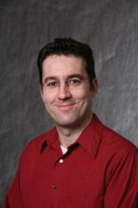 Xavier Intes, an Associate Professor in the Biomedical Engineering Department at Rensselaer Polytechnic Institute, Troy, U.S.. Dr. Intes received his Ph.D. in physics from Université de Bretagne Occidentale, France. He was a postdoctoral fellow at the University of Pennsylvania under the mentorship of Britton Chance and Arjun Yodh. Dr. Intes was the Chief Scientist of Advanced Research Technologies Inc, Montreal, Canada, and oversaw the development of two commercial time-resolved tomographic optical imaging platforms: Optix® and SoftScan®. Dr. Intes’ research interests are on the application of optical techniques for biomedical imaging in pre-clinical and clinical settings. His research concentration is on functional imaging of the breast and brain, fusion with other modalities, and fluorescence molecular imaging. The goal of his laboratory is to develop quantitative thick-tissue optical imaging platforms by focusing on three main areas: (a) design of new optical tomographic imaging instrumentation; (b) developing new reconstruction algorithms for quantitative volumetric imaging; and (c) investigating optimal experimental and theoretical parameters for functional, molecular, and dynamical optical imaging
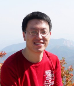 Liang Li, an associate professor at Tsinghua University in Department of Engineering Physics. He received his PhD and Bachelor degrees from Tsinghua University in 2007 and 2002, respectively. His research interests center on the mathematical and physical problems of x-ray imaging and its medical, industrial, and other applications, especially the reconstruction problems under the special imaging conditions. The current projects include: multi-source superfast CT imaging, region-of-interest (ROI) imaging, spectral CT imaging with new photon counting detector, low dose CT reconstruction, dual-modality image reconstruction in MRI&CT, and developments of new medical x-ray imaging devices using digital flat-panel detectors. He has authored 80 peer-reviewed journal and conference papers. He serves as an Editorial Board member for Computerized Tomography Theory and applications, and the leading Guest Editor of Computational and Mathematical Methods in Medicine for the special issue entitled “Mathematical Methods and Applications in Medical Imaging.”
Title 6: Clinical Potential of Noninvasive Bioelectric Imaging
Session organizers: Dana Brooks, Linwei Wang
Thursday, 1 May – 14:00-15:40 – Room R&F3
Session Description:
The theme of this special session is on the current and future clinical potential of a relatively novel technique of quantitative imaging — noninvasive bioelectric imaging. As bioelectricity is invisible in structural images and typically it is not practicable to directly measure tissue bioelectrical signals without invasively interrupting or damaging the tissue, the ability to inversely reconstruct bioelectrical activity in deep tissue — using noninvasive recordings of the electromagnetic fields they produce — could critically improve our ability to assess individualized pathophysiological conditions for individual subjects. In analogy to computed tomographic imaging, these research efforts can be in general seen as computed electrical imaging. They involve acquisition of noninvasive signals external to the body, along with anatomical structures (typically subject specific) and tissue conductivities (typically assumed based on literature studies), followed by an image-based reconstruction algorithm to mathematically and computationally estimate the electrical activity within the organ. It is the goal of this proposed special session to focus on developments in two major areas of noninvasive bioelectric imaging: brain source imaging using electroencephalograpic (EEG) / magnetoencephalographic (MEG), and cardiac source imaging using electrocardiographc (ECG) / magnetocardiographic (MCG) data. We will sample the current state of bioelectric imaging in clinical applications, discuss the implication of these efforts on the future clinical, and commercial, translation of this research, and to identify the accompanying opportunities as well as challenges.
Biography of the organizers:
 Linwei Wang received the B.E. degree in Information engineering from Zhejiang University, Zhejiang, China, in 2005, the M.Phil. degree in electrical and electronic engineering from the Hong Kong University of Science and Technology, Hong Kong, in 2007, and the Ph.D. degree in computing and information science from the Rochester Institute of Technology, Rochester, NY, in 2009. She has been an Assistant Professor in the Ph.D. program of computing and information sciences at the Rochester Institute of Technology since 2009. Dr. Wang is currently directing the Computational Biomedicine Laboratory, and her research interests include computational systems biomedicine, medical image computing, computational cardiac electrophysiology, data-driven statistical inference and computational modeling. She is currently a member of IEEE and American Heart Association.
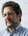 Dana Brooks is Professor of Electrical and Computer Engineering, Associate Director of the Communications and Digital Signal Processing Research Center, co-founder of the Biomedical Signal Processing, Imaging, Reasoning, and Learning (B-SPIRAL) group. and PI of the BioMedical Imaging and Signal Processing Center, all at Northeastern University. He has been a member of the Northeastern ECE faculty since 1991. He is director of the estimation core of the Center for Integrative Biomedical Computing, an NIH/NIGMS P41 Biomedical Technology Research Resource headquartered at University of Utah. Dr. Brooks received the BA in English (’72) from Temple University, and the BSEE (’86), MSEE (’88), and PhD (’91) in Electrical Engineering from Northeastern University. He was a visiting professor during 1999-2000 at the Universitat Polit`ecnica de Catalunya in Barcelona, Spain and a visiting investigator at Memorial Sloan Kettering Cancer Center in fall 2013. His research interests lie in application of statistical and digital signal and image processing to biomedical signal processing and medical and biological imaging, and in open-source software systems for these applications.
Title 7: Applications of Ultrafast Ultrasound Imaging
Session organizers: Hervé Liebgott, Damien Garcia
Friday, 2 May – 11:15-12:55 – Room R&F3
Session Description:
Medical ultrasound imaging is currently going through a real revolution with the growing emergence of ultrafast ultrasound imaging. Whereas ultrasound imaging was still limited to frame rates of hundred of Hertz a decade ago, ultrafast ultrasound imaging is nowadays able to produce up to 15.000 images per second. This offers new perspectives especially when motion estimation becomes of interest. The special session that we propose aims at giving an overview of one of the hottest topics in medical ultrasound.
Biography of the organizers:
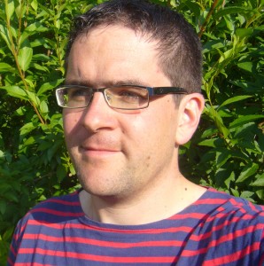 Hervé Liebgott is associate professor at the University of Lyon in the CREATIS Laboratory. His research focuses on image and signal processing applied to medical ultrasound imaging. He is particularly interested in image formation techniques and motion estimation. He received the PhD degree in 2005 for his research on Transverse Oscillations techniques adapted to elastography. His has been a visiting PhD student at the Technical University of Denmark for 6 months between 2003 and 2005 and he is currently and for 6 months an invited professor at the University of Montreal. In 2011 he received the French Habilitation to Lead Research (HDR) from University Lyon 1. He is currently the vice leader of the Ultrasound imaging team at CREATIS. He became in 2014 an Associate member of the Bio Imaging and Signal Processing (BISP) technical committee of the IEEE Signal Processing Society.
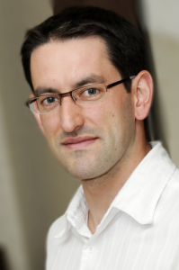 Damien Garcia was born in 1974 in Paris, France. He obtained the DEA and engineer degree of Centrale Marseille in 1997, and the M.Sc. and Ph.D. degrees in biomedical engineering from the University of Montreal in 2003. He was a postdoctoral fellow in 2006-2008 at the department of echocardiography, Gregorio Marañón hospital, Madrid, Spain. Dr. Garcia is director of the Research Unit of Biomechanics & Imaging in Cardiology (RUBIC) at the University of Montreal Hospital Research Centre (CRCHUM), and assistant professor at the department of radiology, radio-oncology and nuclear medicine at the University of Montreal. His research interests are in cardiovascular ultrasound imaging, Doppler echocardiography, image processing, mathematical and biomechanical modeling, fluid dynamics and flow imaging. Damien Garcia holds a research scholarship from the Fonds de Recherche en Santé du Québec (FRSQ). A detailed list of his publications is available in www.biomecardio.com.

