
Biomedical image registration
Wednesday, April 14, 08:30 - 12:15
Presented by
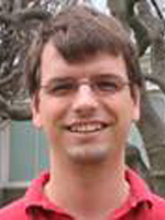
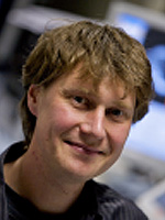
Presented by Gustavo Rohde (Carnegie Mellon University, USA) and Graeme Penney (King's College London, UK)
Description
Part I: Overview of Principles (Gustavo K. Rohde)
Digital image registration has been a topic of study in engineering related fields for several decades. Many important ideas have been introduced, developed, tested, and applied with great success especially in applications related to biomedical imaging. This portion of the tutorial is designed to introduce some of the most important principles used in modern solutions to image alignment problems. It will be focused on the ideas that have been particularly useful for biomedical imaging applications. Familiarity with basic signal and image processing techniques (e.g. interpolation, optimization, sampling, Fourier analysis, etc.) will be assumed.
Problem Statement
- Preliminaries: image data, applications, spatial transformations
- Registration as an estimation problem
Point-Based Registration
- Points, landmarks, features
- Procrustes problem
- Nonrigid transformations using radial basis functions
Intensity-Based Registration
- Phase correlation
- Objective functions (least squares, cross correlation, mutual information, multi-channel)
- Numerical optimization methods: parametric, non-parametric, small vs. large deformation
- Uncertainty estimates
Metric Image Registration
- Metrics: metrics on diffeomorphisms, optimal transportation
Registration in Quantitative Imaging
- Effects of interpolation, noise, uncertainty
- Applications in motion and distortion correction, morphometry
About the Speaker
Gustavo K. Rohde received the B.S. degrees in physics and mathematics in 1999 and the M.S. degree in electrical engineering in 2001 from Vanderbilt University, Nashville, TN. He received the Ph.D. degree in applied mathematics and scientific computation in 2005 from the University of Maryland, College Park. He was a research assistant in a biomedical imaging laboratory at the National Institutes of Health, USA, from 2001 to 2005, and a National Research Council postdoctoral associate at the Naval Research Laboratory, USA, in 2005 and 2006. Since 2007, he has been an Assistant Professor of biomedical engineering and electrical and computer engineering (by courtesy), and a faculty member of the Lane Center for Computational Biology, at Carnegie Mellon University, Pittsburgh, PA, where he is also a core faculty member of the Center for Bioimage Informatics. His research interests include the application of image processing and pattern recognition techniques to various biomedical problems.
Part II: Applications in Image Guided Interventions (Graeme P. Penney)
One of the main application areas for image registration has been in the fields of image-guided surgery and interventions. The major clinical benefits from minimising healthy tissue damage, has led surgeons to investigate methods to help guide them during operations. This is particularly important where the surrounding tissue is largely non-regenerative, such as in neurosurgery. The minimally invasive revolution has led to clinicians requiring new methods to observe the surgical scene for a wide range of applications. There has been a merging of the skills between interventional radiologists and surgeons, as therapy has been guided solely by interventional images such as fluoroscopy, endoscopy or ultrasound, with no direct line of sight to the surgical scene. Image registration offers the potential to enhance the clinicians visualisation during these procedures, by aligning higher quality diagnostic images to the interventional images. Part II of this tutorial will describe how image registration technologies are used to aid surgical guidance. This tutorial will cover a wide range of applications and methods will be covered, from those in widespread clinical use, to those which are currently being proposed in what is a very active research area. Continuing improvements in computational power and image quality are increasingly making feasible the applications of new image guidance algorithms to wider range of clinical procedures.
History of Image Guided Interventions
Components of an Image Guidance System
- User specifications for IGI
- Tracking Systems
- Registration methods
- Visualisation
Registrations to a Physical Coordinate System
- Applications in neuro and orthopaedic surgery
- Extensions to applications involving soft tissue structures
Registrations to Interventional Images
- Projective interventional images: fluoroscopy and optical
- Applications in cardiac and vascular interventions and radiotherapy
- Alignment to ultrasound images
- Applications in cardiac and abdominal interventions
Future Developments
- Non-rigid developments
- Synergy with robotics
About the Speaker
Graeme P. Penney received his BSc in Physics from the University of Durham in 1992, and his PhD from King's College London in 2000, working on 2D-3D image registration techniques. He spent a year in the Image Sciences Institute, Utrecht working on image interpolation techniques and two years at University College London working on ultrasound registration. He has just completed an EPSRC Advanced Research Fellowship and is now a Senior Lecturer in Image Processing at King's College London. His main research interests are on developing techniques to register preoperative images to interventional images to enable preoperative information to be used during interventional procedures. He is author of one of the most widely cited papers on 2D-3D medical images registration, and has over sixty refereed journal/conferences publications on medical image registration methods.