Tutorial T-4: Optics for In Vivo Imaging and Monitoring in Biology and Medicine
Wednesday, March 30, 08:30 - 12:15
Presented by
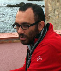
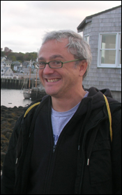
Turgut Durduran (ICFO-The Institute of Photonic Sciences, Castelldefels, Spain) and Jorge Ripoll (Institute for Electronic Structure and Laser – FORTH, Greece)
Description
Part I: Fundamentals
A) Fundamentals of Optics in BioMedicine (Jorge Ripoll)
In this first fundamental section, the origins of color in nature will be explained based on light emission, light absorption and light scattering. Within these categories, we will explain how scattering and absorption affects light propagation, and introduce the basics of light emission including bioluminescence, fluorescence and laser. All these effects will be discussed within the context of light propagation in tissues, and will include a description of the main absorbing and scattering components present when imaging in-vivo.
B) Fundamentals of Tissue Spectroscopy and Imaging (Turgut Durduran)
Tissue spectroscopy and imaging refers to a wide-range of optical modalities and applications. In these tutorials, I focus on those that utilize near-infrared (~650-950nm) which are the most suitable for “deep-tissue” (~cm) spectroscopy and imaging (tomography). I start with a brief outline of non-quantitative studies going back to 1920s (“optical mammography”) and continue with early quantitative studies on brain (“functional near-infrared spectroscopy” (fNIRS)) and the development of the pulse-oximeter which is widely utilized in the clinics. Finally, these will lead way to the fundamentals of “modern” near-infrared spectroscopy (NIRS) and diffuse optical tomography (DOT).
Part II: Applications
A) Applications in Biology (Jorge Ripoll)
Once the main governing principles of light propagation have been covered in the first introductory part, we shall here address how the amount of scattering present in a sample dictates the different imaging modality to be employed. To that end, we shall cover several applications in biology depending on the scattering properties of the tissue, namely, samples where light propagation is ballistic and samples where light propagation is diffusive. With these applications in mind, we shall discuss new imaging techniques such as Optical Projection Tomography (OPT) and Selective Plane Illumination Microscopy (SPIM) as ballistic imaging approaches, and Fluorescence Molecular Tomography (FMT) and related techniques as diffuse imaging approaches. The issue of improving image resolution in highly scattering media will be discussed.
B) Applications on Humans in Clinical Settings (Turgut Durduran)
This part of the tutorial will focus on studies on humans in clinical settings. An attractive feature of diffuse optical technologies is that they could be utilized in the clinics without posing any major safety issues in populations ranging from premature born infants to critically ill adults. By allowing non-invasive monitoring and imaging of microvascular blood flow, blood oxygenation and blood volume, they could help both in diagnosis and in therapy guidance. I will show examples from “traditional” methods in cancer therapy and in neuro-intensive care monitoring. Some relatively newer approaches of fluorescence tomography and diffuse correlation spectroscopy will also be demonstrated.
About the Speakers

Turgut Durduran
received his Bs. A. and Ph.D. degrees in physics at University of Pennsylvania (UPENN), USA. He has then spent four more years at UPENN as a researcher with a dual appointment in Departments of Physics & Astronomy and Radiology. In 2009, he moved to ICFO-The Institute of Photonic sciences in Castelldefels, Barcelona, Spain where he leads a research group on medical optics. His research interests are mainly focused on the clinical translation of diffuse optical technologies in neurology and oncology and the development of diffuse correlation spectroscopy (DCS).
Jorge Ripoll
received the B.S. degree and Ph.D. in physics at the Universidad Autonoma of Madrid (UAM), Spain in 2000. He has been lucky enough to have the opportunity to work in several labs both in Europe and the USA including UCL, UPENN and Harvard. He is currently a visiting Professor at the ETH in Zurich, and since 2005 holds a permanent position at the Foundation for Research and Technology-Hellas (FORTH) where he leads a research group on in-vivo optical imaging. His research interests are in-vivo optical imaging applications in biology, mainly new 3D microscopy and whole animal imaging approaches.