Tutorial T-1: Photoacoustic Tomography: Ultrasonically Breaking through the Optical Diffusion Limit
Wednesday, March 30, 08:30 - 12:15
Presented by
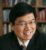
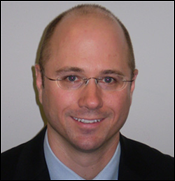
Lihong Wang and Mark Anastasio, Department of Biomedical Engineering, Washington University in St. Louis, USA
Description
Part I: Principles and Applications (Lihong V. Wang)
Photoacoustic tomography has been developed for in vivo early-cancer detection and functional imaging by physically combining non-ionizing electromagnetic and ultrasonic waves. Unlike ionizing x-ray radiation, non-ionizing electromagnetic waves—such as optical and radio waves—pose no health hazard and reveal new contrast mechanisms. Unfortunately, electromagnetic waves in the non-ionizing spectral region do not penetrate biological tissue in straight paths as x-rays do. Consequently, high-resolution tomography based on non-ionizing electromagnetic waves alone—such as confocal microscopy, two-photon microscopy, and optical coherence tomography—is limited to superficial imaging within approximately one optical transport mean free path (~1 mm in the skin) of the surface of scattering biological tissue. Ultrasonic imaging, on the contrary, provides good image resolution but has strong speckle artifacts as well as poor contrast in early-stage tumors. Ultrasound-mediated imaging modalities that combine electromagnetic and ultrasonic waves can synergistically overcome the above limitations. The hybrid modalities provide relatively deep penetration at high ultrasonic resolution and yield speckle-free images with high electromagnetic contrast.
In photoacoustic computed tomography, a pulsed broad laser beam illuminates the biological tissue to generate a small but rapid temperature rise, which leads to emission of ultrasonic waves due to thermoelastic expansion. The short-wavelength pulsed ultrasonic waves are then detected by unfocused ultrasonic transducers. High-resolution tomographic images of optical contrast are then formed through image reconstruction. Endogenous optical contrast can be used to quantify the concentration of total hemoglobin, the oxygen saturation of hemoglobin, and the concentration of melanin. Melanoma and other tumors have been imaged in vivo. Exogenous optical contrast can be used to provide molecular imaging and reporter gene imaging.
In photoacoustic microscopy, a pulsed laser beam is focused into the biological tissue to generate ultrasonic waves, which are then detected with a focused ultrasonic transducer to form a depth resolved 1D image. Raster scanning yields 3D high-resolution tomographic images. Super-depths beyond the optical diffusion limit have been reached with high spatial resolution.
Thermoacoustic tomography is similar to photoacoustic tomography except that low-energy microwave pulses, instead of laser pulses, are used. Although long-wavelength microwaves diffract rapidly, the short-wavelength microwave-induced ultrasonic waves provide high spatial resolution, which breaks through the microwave diffraction limit. Microwave contrast measures the concentrations of water and ions.
The annual conference on this topic has been doubling in size approximately every three years since 2003 and has become the largest in SPIE’s Photonics West as of 2009.
- Introduction
- Motivation for optical imaging
- Diffraction and diffusion limits
- Photoacoustics
- Signal generation
- Photoacoustic wave equation
- Photoacoustic Doppler effect
- Photoacoustic computed tomography
- Circular geometry
- Linear geometry
- Photoacoustic microscopy
- Acoustic resolution
- Optical resolution
- Discussion and summary
- Thermoacoustic tomography
- Multiscale photoacoustic tomography
- Summary of pros and cons
About the Speaker

Lihong V. Wang
earned his Ph.D. degree at Rice University, Houston, Texas under the tutelage of Robert Curl, Richard Smalley, and Frank Tittel and currently holds the Gene K. Beare Distinguished Professorship of Biomedical Engineering at Washington University in St. Louis. His book entitled “Biomedical Optics: Principles and Imaging,” one of the first textbooks in the field, won the 2010 Joseph W. Goodman Book Writing Award. He also coauthored a book on polarization and edited the first book on photoacoustic tomography. He serves as the Editor-in-Chief of the Journal of Biomedical Optics. Professor Wang has published >250 peer-reviewed journal articles and delivered >270 keynote, plenary, or invited talks. He is a Fellow of the AIMBE (American Institute for Medical and Biological Engineering), the OSA (Optical Society of America), the IEEE (Institute of Electrical and Electronics Engineers), and the SPIE (Society of Photo-Optical Instrumentation Engineers). He chairs the annual conference on Photons plus Ultrasound, and chaired the 2010 Gordon Conference on Lasers in Medicine and Biology and the 2010 OSA Topical Meeting on Biomedical Optics. He is a chartered member on an NIH Study Section. Wang is the founding chairs of the scientific advisory boards for two companies commercializing his inventions. He received FIRST and CAREER awards. He has received 27 research grants as the principal investigator with a cumulative budget of >$30M. His laboratory invented or discovered the functional photoacoustic tomography, dark-field confocal photoacoustic microscopy (PAM), optical-resolution PAM, photoacoustic Doppler effect, photoacoustic reporter gene imaging, focused scanning microwave-induced thermoacoustic tomography, the universal photoacoustic or thermoacoustic reconstruction algorithm, frequency-swept ultrasound-modulated optical tomography, time-reversed ultrasonically encoded (TRUE) optical focusing, sonoluminescence tomography, Mueller-matrix optical coherence tomography, optical coherence computed tomography, and oblique-incidence reflectometry. In particular, PAM broke through the long-standing diffusion limit to the penetration of conventional optical microscopy and reached super-depths for noninvasive biochemical, functional, and molecular imaging in living tissue at high resolution. Professor Wang’s Monte Carlo model of photon transport in scattering media is used worldwide.Part II: Image Reconstruction (Mark A. Anastasio)
Photoacoustic computed tomography (PCT) is a computed imaging modality that utilizes an image reconstruction algorithm to form an image that depicts the absorbed optical energy distribution. A variety of image reconstruction algorithms have been developed for three-dimensional PCT. All known analytic reconstruction algorithms that are mathematically exact and numerically stable require complete knowledge of the photoacoustic wavefield on a measurement aperture that either encloses the entire object or extends to infinity. In many potential applications of PCT it is not feasible to acquire such measurement data. Because of this, iterative, or more generally, optimization-based, reconstruction algorithms for PCT are being developed actively that provide the opportunity for accurate image reconstruction from incomplete measurement data. Iterative reconstruction algorithms also allow for the use of accurate imaging models that account for the response of the measurement system and the relevant wave physics.
In this component of the tutorial we review the principles of PCT image reconstruction. This will include a comprehensive description of imaging models and the associated image reconstruction algorithms.
- Imaging physics
- Photoacoustic wave equation
- Frequency-dependent acoustic attentuation
- Effects of heterogeneties in acoustic properties
- Ultrasound transducer modeling
- Basic principles of PCT image reconstruction
- Canonical PCT imaging model in its continuous form
- The Fourier-shell identify
- Spatial resolution from a Fourier perspective
- Analytic image reconstruction methods
- Data symmetries and redundancies
- Identification of data redundancies
- Half-time image reconstruction
- Half-scan image reconstruction
- Methods for iterative image reconstruction
- Continuous-to-discrete PCT imaging models
- Finite-dimensional object representations
- Discrete-to-discrete PCT imaging models
- Formulation of optimization problem
About the Speaker

Mark A. Anastasio
earned his Ph.D. degree at the University of Chicago in 2001 and is currently a Professor of Biomedical Engineering at Washington University in St. Louis (WUSTL). Prior to joining WUSTL, he was a faculty member at Illinois Institute of Technology. Dr. Anastasio is an internationally recognized expert on tomographic image reconstruction, imaging physics, and the development of novel biomedical imaging systems. He has conducted pioneering research in the fields of diffraction tomography, X-ray phase-contrast imaging, and photoacoustic tomography. He has published over 60 peer review articles on these topics and was a recipient of an NSF CAREER award. He is on the Editorial Board of the Journal of Biomedical Optics and is on the organizing committee for the SPIE Photonics West Photons plus Ultrasound conference and the SPIE Conference on Image Reconstruction from Incomplete data. He was also a Program Chair for the 2009 Optical Society of America Signal Recovery and Synthesis Topical Meeting.